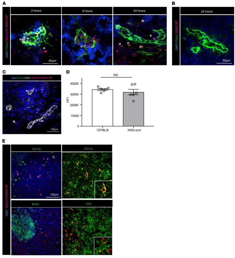
Figure 2. Histological characterization of MECA79-NP trafficking in the DLN.
(A) MECA79-IR800-NPs (in red) can be detected in the HEV (in green) at early time points and infiltrate to adjacent areas in parenchyma of LNs at later time points. (B) Nontargeted IR800-NPs were not detected in the DLN. (C) Lack of PNAd expression and impaired trafficking of MECA79-IR800-NPs to the LNs of double-knockout mice. (D) MFI quantification of axillary LNs showing no difference between C57BL/6 mice and NOD-scid mice at 24 hours after injection of MECA79-IR800-NPs (34,356 ± 837.7 vs. 31,918 ± 2,515, mean ± SEM, Student’s t test, P = NS, n = 3 mice per group, 2 LNs from each mouse). (E) Immunofluorescence analysis of the DLN showed colocalization of MECA79-IR800-NPs with CD11c+ and CD3+ cells (scale bar: 100 μm).

