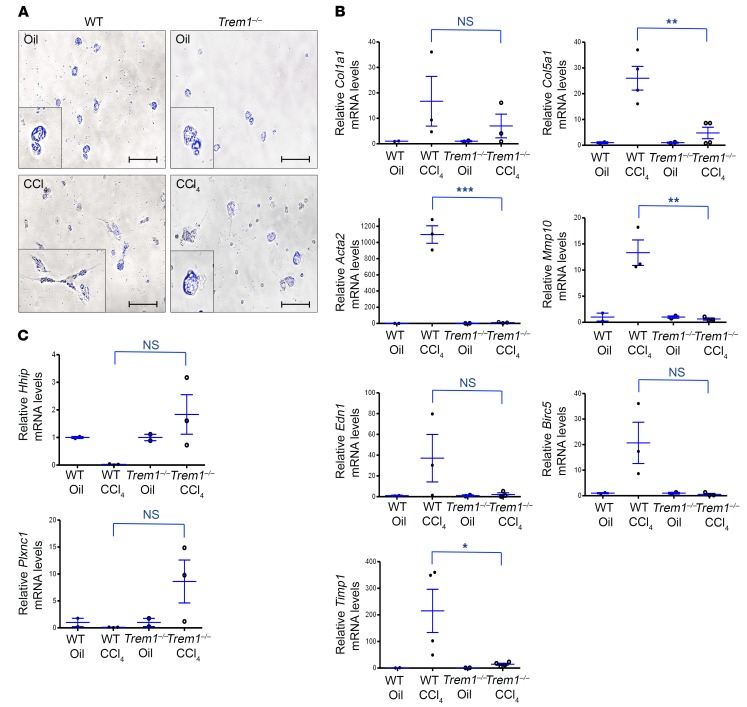
Figure 2. Deletion of Trem1 attenuates HSC activation and differentiation.
HSCs were isolated from oil-injected (Oil) control WT and Trem1–/– mice (n = 3/group) and from WT and Trem1–/– mice treated with CCl4 for 6 weeks (n = 3–4/group). (A) Representative images of freshly isolated HSCs from the indicated mice (oil-injected, top; CCl4-treated, bottom), visualized using a merging of phase-contrast microscopy and retinoid fluorescence (blue channel), show that HSCs from WT CCl4-injured mice differentiated into myofibroblasts and lost their retinoic acid droplets. Original magnification, ×40; scale bars: 25 μm. Images shown are representative of 2 independent experiments. (B and C) Total RNA was isolated from HSC fractions from WT or Trem1–/– mice treated with CCl4 for 6 weeks (n = 3–4/group). Col1a1, Col5a1, Acta2, Mmp10, Edn1, Birc5, Timp1, Hhip, and Plxnc1 mRNA levels were determined by RT-qPCR and are represented as the fold induction. Results are displayed as the mean ± SEM. *P < 0.05, **P < 0.01, and ***P < 0.001, by 2-tailed Student’s t test.

