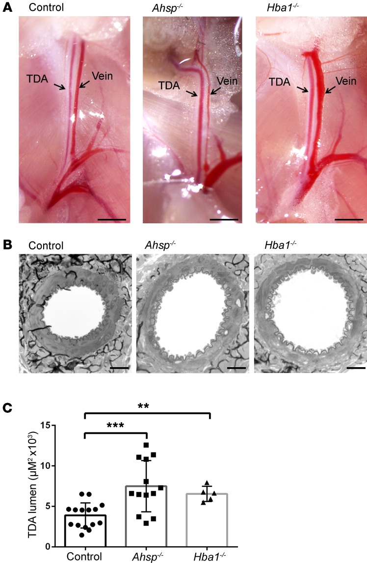
Figure 3. Ahsp or Hba1 gene disruption causes steady-state TDA dilation in vivo.
(A) TDAs and veins in situ. Scale bars: 5 mm. (B) Electron micrographs of TDA cross sections from 24-week-old mice. Scale bars: 20 μm. (C) Histomorphometric analysis of randomly sampled TDA cross sections from WT control mice (n = 5, 15 random measurements), Ahsp−/− mice (n = 5, 13 random measurements), and Hba1−/− mice (n = 4, 5 random measurements). **P < 0.01 and ***P < 0.005, by unpaired t test.

