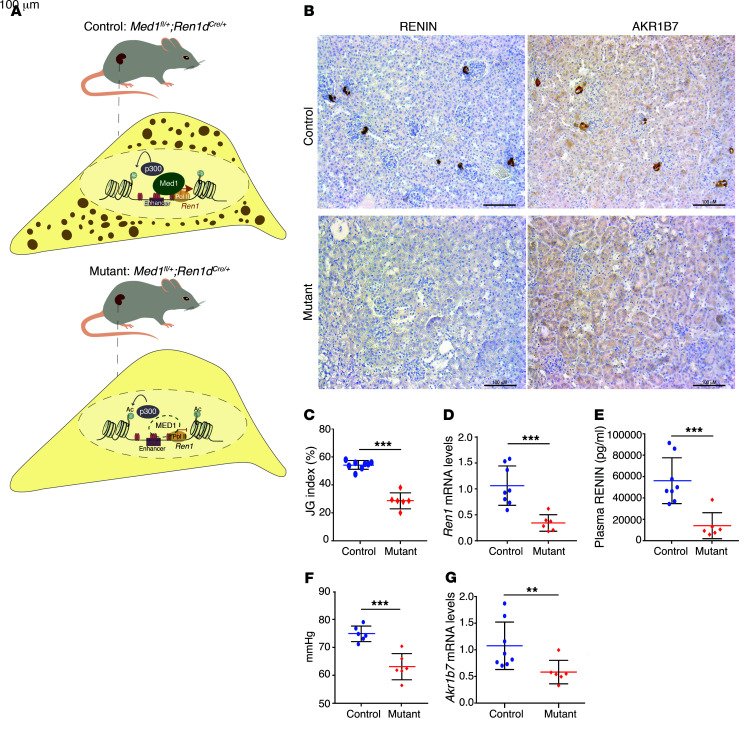
Figure 6. The coactivator MED1 is essential for renin expression in vivo.
(A) Control mouse where one allele of Med1 gene is floxed and the other is WT. The cell below indicates that in the presence of Med1 the renin gene is transcribed and there is granule formation and renin storage. Mutated mouse where one allele of Med1 is floxed and the other one is deleted; the Cre gene driven by the renin promoter deletes the floxed allele, generating a Med1 homozygous deletion in renin lineage cells. In the cell corresponding to the mutant mouse, absence of MED1 (green dashed line) indicates that there is no renin transcription and no granule formation in the renin cells. (B) Immunostaining for RENIN and AKR1B7 shows a decrease in signal for both proteins in JG cells of mutant mice (n = 6) compared with control mice (signal is indicated by the brown color; n = 8). (C) The JG index, measured as the percentage of RENIN-positive areas over the total number of glomeruli, indicates that mutant mice (n = 6) have fewer renin-positive JG areas when compared with control mice (n = 8). (D) Relative expression of renin mRNA levels measured by qPCR. Mutant mice (n = 6) have significantly lower expression than the controls (n = 8). (E) Plasma RENIN levels are lower in mutant mice (n = 6) than in controls (n = 8). (F) Med1 mutant mice have lower blood pressures than control mice (n = 6 per group). (G) Akr1b7 mRNA levels are decreased in mutant mice (n = 6). Data are mean ± SD. **P < 0.01; ***P < 0. 001, by unpaired, 2-sided Student’s t test.

