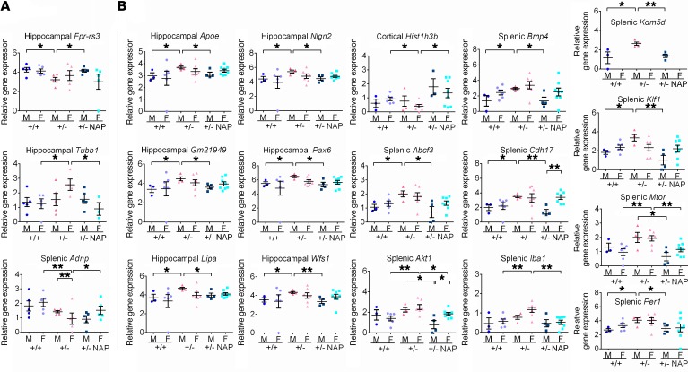
Figure 4. The Adnp genotype affects gene expression in the brain and spleen in 19- and 27-day-old and 3-month-old mice, with significant amelioration following NAP treatment.
HT qRT-PCR was performed on mRNA extracted from hippocampus, cortex, and spleens of 19- to 27-day-old mice (males: Adnp+/+ n = 5, Adnp+/– n = 5, Adnp+/– NAP, n = 5; females: Adnp+/+ n = 6, Adnp+/– n = 4, Adnp+/– NAP, n = 5) and of 3-month-old male (M) and female (F) mice (males: Adnp+/+ n = 3, Adnp+/– n = 4, Adnp+/– NAP, n = 4; females: Adnp+/+ n = 6, Adnp+/– n = 7, Adnp+/– NAP, n = 8). Results were normalized to Hprt. Significantly affected genes in 19- to 27-day-old mice (A) and 3-month-old mice (B) are presented. An unpaired Student’s t test revealed significant differences between vehicle-treated Adnp+/+ and Adnp+/– mice and between NAP- and vehicle-treated Adnp+/–mice (*P < 0.05 and **P < 0.01). Additional Student’s t tests were performed, comparing data between male and female mice to determine sex differences. All reported P values were also significant after multiple comparisons correction at a FDR of 10%.

