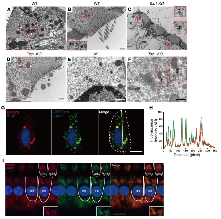
Figure 10. Peroxisomes are involved in the regulation of mTORC1 in inner ear hair cells.
(A–F) TEM analysis revealing the morphologies of mitochondria and peroxisomes in cochlear OHCs of WT and Tsc1-cKO mice at P28. (A and D) No mitochondrial abnormalities were seen at this time point in Tsc1-cKO mice compared with WT controls. The mitochondria showed normal morphologies with defined lamellar cristae. (B, C, E, F) Normally, peroxisomes (red arrowheads) demonstrate a circular cytoplasmic arrangement around a central nucleus in WT mice (B and E). However, many crystalline nuclei appeared in the peroxisomes of Tsc1-cKO OHCs (C), and several abnormal peroxisomes had no evident membrane structures (F). Scale bars: 1 μm (A–C), 0.5 μm (D–F). (G) Tsc1 is associated with peroxisomes in transfected HEI-OC1 cells. Transfected HEI-OC1 cells producing Tsc1 (Tsc1-GFP, green) were coimmunostained with an antibody against PMP70 (red). Cell nuclei were stained with DAPI (blue). Scale bar: 10 μm. (H) Corresponding line intensity measurements for each protein indicate strong colocalization of GFP (green) and PMP70 in peroxisomes. The fluorescence intensity was quantified using ImageJ (NIH). (I) Double immunolabeling of hair cells (IHCs and OHCs) for PMP70 (red) and endogenous Tsc1 (green). Scale bar: 10 μm. Three mice were examined for each genotype.

