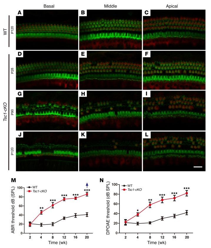
Figure 6. Tsc1-cKO mice undergo hair cell degeneration and gradual hearing loss from the fourth week of life.
Confocal images of cochlear whole mounts labeled with a Myo7A antibody (red) and counterstained with phalloidin to label filamentous actin (green) are shown. (A–C) Representative images from the basal, middle, and apical turns of the organ of Corti in WT mice at P120. (D–L) Representative images of the organ of Corti from 3 turns of the cochleae in Tsc1-cKO mice at P28 (D–F), P90 (G–I), and P120 (J–L). OHCs began to degenerate by P28. Severe degeneration was evident at P90. By P120, only a few OHCs and partial IHCs remained in the middle and apical turns of the cochleae. n = 5 for each group. (M and N) Graphs illustrate age-dependent threshold changes in WT and Tsc1-cKO mice. Age-related click ABR thresholds (M) and age-related DPOAE thresholds (N) in WT and Tsc1-cKO mice showed progressive hearing loss. The group sizes were as follows: 2 and 4 weeks: n = 15 for each group; 8, 12, 16, and 20 weeks: n = 5 for each group. The arrow indicates that even at the highest SPL test level (90 dB SPL), several Tsc1-cKO mice showed no response. Data are shown as the mean ± SD. *P < 0.05, **P < 0.01, ***P < 0.001, by 2-tailed Student’s t test. Scale bar: 20 μm.

