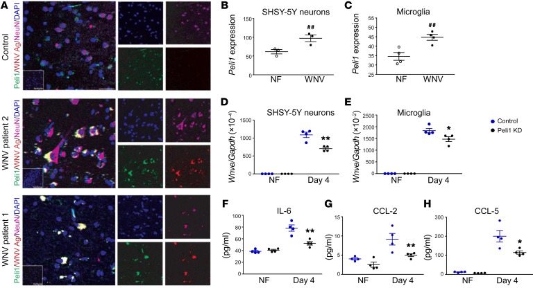
Figure 6. Peli1 expression and its role in human cells during WNV infection.
(A) Immunodetection of Peli1 (green), WNV antigen (WNV Ag) (red), and NeuN (purple), in postmortem hippocampal tissues from 1 control and 2 WNV-infected patients. Nuclei were counterstained with DAPI (blue). Scale bar: 8 μm. Insets are images of sections stained with isotype control antibodies or serum (original magnification, ×63). (B and C) RNA levels of Peli1 in SH-SY5–derived neurons and human microglia on days 1 and 4 p.i. were determined by qPCR. Data are presented as the mean ± SEM and are representative of 2 similar experiments (n = 3–4). ##P < 0.01 compared with the noninfected group (unpaired, 2-tailed Student’s t test). (D–H) SHSY-5Y–derived neurons (D) and human microglial cells (E–H) were treated with control or Peli1 siRNA (Peli1 KD), infected with WNV 385-99 at 48 hours, and harvested at the indicated time points. (D and E) Viral load was measured on day 4 by qPCR. (F–H) IL-6, CCL-2, and CCL-5 production in microglial cells was measured on day 4 by Bio-Plex assay. Data are presented as the mean ± SEM and are representative of 2 similar experiments (n = 4). (D–H) *P < 0.05 and **P < 0.01 compared with the control group (unpaired, 2-tailed Student’s t test).

