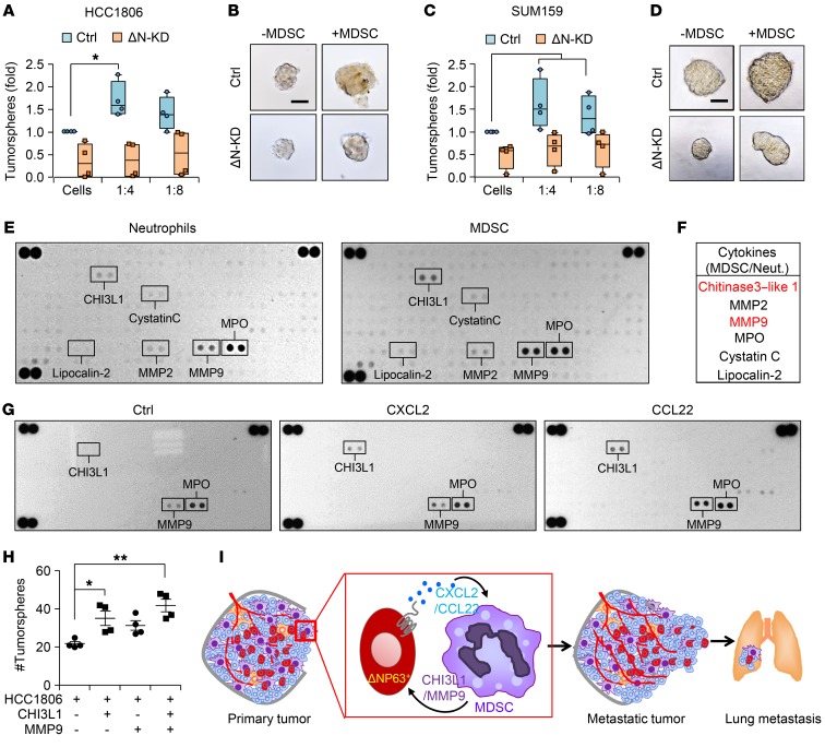
Figure 7. MDSCs secrete prometastatic factors and enhance CSC activities of TNBC.
(A and B) Number (A) and representative images (B) of tumorspheres from HCC1806 cells cultured for 3 days with or without MDSCs. (C and D) Number (C) and representative images (D) of tumorspheres from SUM159 cells cultured for 3 days with or without MDSCs. (A–D) n = 3 independent experiments performed in technical duplicate. (E) Representative images of cytokine arrays from cell lysates of normal mammary neutrophils (from mammary gland) and MDSCs from primary mammary tumor. (F) Table shows the most differentially expressed proteins. (G) Representative array blots show differential protein expression upon treatment of MDSCs with recombinant CXCL2 and CCL22 for 12 hours. (H) Scatter plot shows number of tumorspheres from HCC1806 cells upon indicated treatments. Scatter plot show data from 3 independent experiments, and data are presented as the mean ± SEM. (I) Model shows the recruitment of PMN-MDSCs at primary tumor and at metastatic organ via ΔNp63+ cancer cells through chemokines. Concomitantly, MDSCs secrete factors such as chitinase 3–like 1 (CHI3L1) and MMP9 to enhance both tumor growth and metastatic potential. Scale bars: 40 μm (B and D). Data are presented as the mean ± SEM. *P < 0.05; **P < 0.01. P value was calculated using 1-way ANOVA with Tukey’s multiple-comparisons post hoc test.

