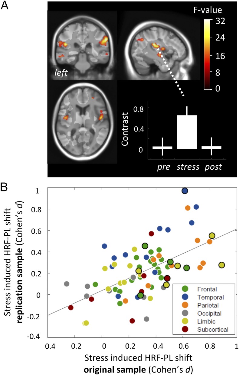
Fig. 7.
Overview on replication results. (A) Voxel-based analysis with statistical map of F-value contrast comparing prestress and stress conditions in the replication sample, showing equally directed and reversible HRF-PL increases in bilateral insular, superior temporal, parietolateral, and right orbitofrontal cortices (collection threshold P < 0.001, six significant whole-brain corrected clusters, Pcluster.FWE < 0.05; for details see SI Appendix, Table S4). (B) Correlation of regional HRF-PL shift effects between samples, calculated from voxels with sufficient latency-corrected BOLD amplitude responses in individual anatomical parcellations (r = 0.585, P = 1.9e-08). Black circles denote atlas regions that showed an HRF-PL shift in the SPM analysis in the original sample (Table 1).

