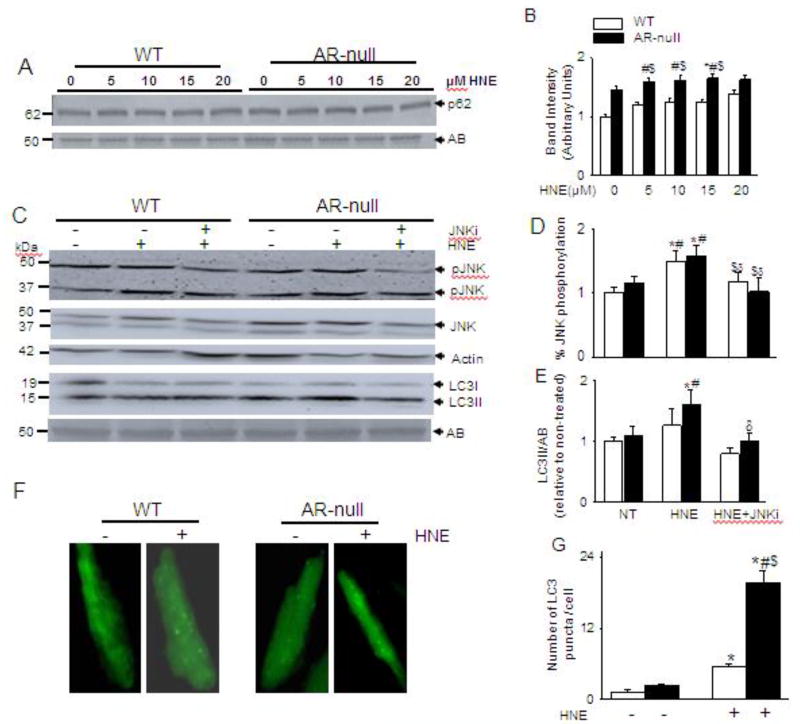
Fig. 6. HNE-induced increase in autophagy is dependent on P62 and JNK.
Isolated cardiac myocytes from WT and AR-null mice were superfused with varying concentrations of HNE (5–20 µM) for 40 min and analyzed for (A) p62 levels. (C) Cardiac myocytes isolated from WT and AR-null cardiac myocytes were pretreated with or or without JNK inhibitor SP 600125 (25 µM for 30 min) and then superfused with HNE (15 µM) for 40 min. Cell lysate were immunoblotted with (C) pJNK and LC3 antibodies. (F) Representative images of isolated adult cardiac myocytes from WT and AR-null hearts transduced with Ad-GFP-LC3 and then perfused with 15 µM HNE for 60 min. (B, D, E,G) Data are mean ± SEM. *p < 0.05 vs non-treated WT, #p < 0.05 vs non-treated AR-null, $ p < 0.01 vs HNE-treated WT cardiac myocytes, δp<0.05 vs AR-null HNE treated cardiac myocytes, n=3–4 mice in each group.

