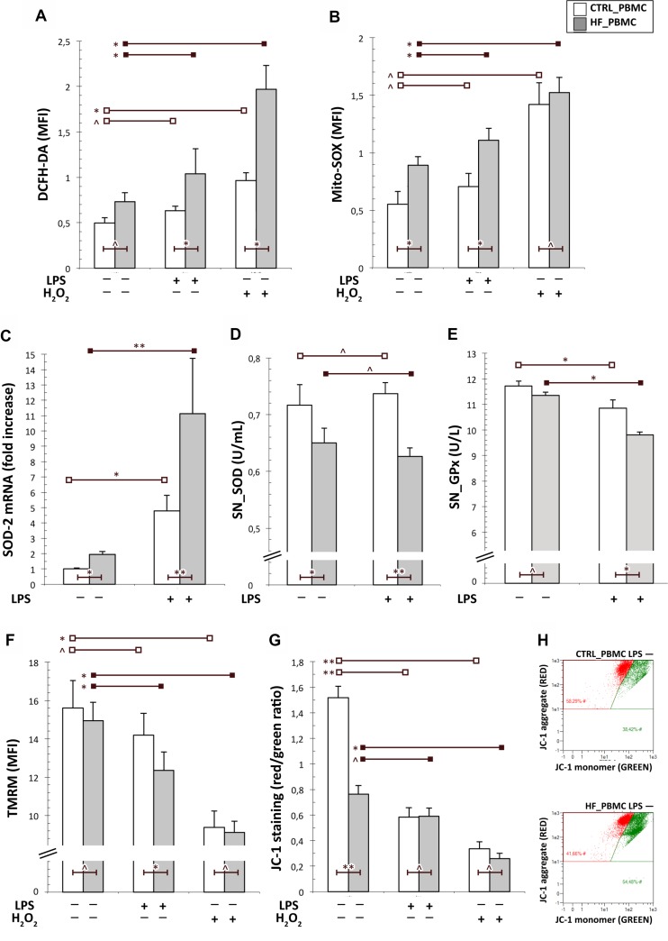
Figure 1. Analysis of the mitochondrial function in PBMCs from HF and CTRL subjects.
(A, B) Cytofluorimetric assay of cytoplasmic and mitochondrial ROS generation in unstimulated or stimulated PBMCs with LPS. The mitochondrial ROS levels were higher in PBMCs from HF respect to CTRL both at baseline and after LPS stimulation. As expected, the stimulation with H2O2 increased both cytoplasmic and mithocondrial oxidative stress particularly in HF_PBMCs. (C) A significant upregulation of the SOD-2 mRNA levels, as detected by RT-PCR, was found in HF_PBMCs with respect to CTRL_PBMCs, particularly after LPS stimulation (Mann–Whitney test: *p < 0.05, **p < 0.01 and ^p = NS). (D, E) Assessment of antioxidant enzymes activity in supernatants of PBMCs cultures; the SOD and GPx activities were decreased in the HF_PBMCs compared to CTRL_PBMC (Student T test: *p < 0.05, **p < 0.01 and ^p = NS). (F–H) Cytofluorimetric assay for mitochondrial membrane potential (Δψm; TMRM staining) and mitochondrial depolarization index (JC-1 staining) in unstimulated or stimulated PBMCs with LPS. The data reflect a significant mitochondrial depolarization in HF_ with respect to CTRL_PBMCs. (Student T test: *p < 0.05, **p < 0.01 and ^p = NS). The panel G shows a JC-1 staining plot from representative PBMCs samples.

