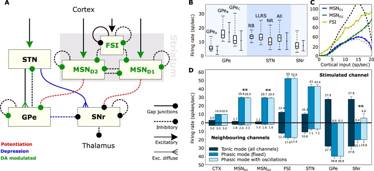
Fig 1. Architecture and firing behaviour of the system.
A: The BG circuit as realized in the present study. Dopamine (DA) influences both the internal behaviour of MSNs and FSIs as well as the impact of various synaptic conductances. B: Firing rates of the various neuron types when isolated. Boxplots show median, first-third quartiles, and minimum-maximum of the mean firing rate of each simulated neuron. Solid error bars represent mean and standard deviation of recorded (real) spontaneous activity. Data for GPe neurons were taken from [30], for STN neurons from [31] and for SNr neurons from [32]. Neuron sub-types of the striatum include medium spiny-projection neurons (MSN) and fast-spiking interneurons (FSI), the STN includes rebound-bursting (RB), long-lasting rebound spikes (LLRS) and no-rebound (NR) neurons, and the GPe includes the three types GPeA−C. C: Cortical input-firing rate curve of striatal neurons when the complete model is in use. The dashed lines illustrate the MSND1 curve for low dopamine (grey) and high dopamine (black) in the system. D: The mean firing rates, over 500 3-second trials, of the various neuron types when the complete model is in use. The stimulated channel represents the channel that received enhanced cortical input during the phasic mode, while the set of bar charts below show the firing rates of the two neighbouring channels. The error bars show standard deviation. In tonic mode, there is no discrimination between channels and the small differences in the two sets of bar charts are the result of random noise. The double asterisk (**) denotes an independent two-sample T-test with p-value < < 0.01.

