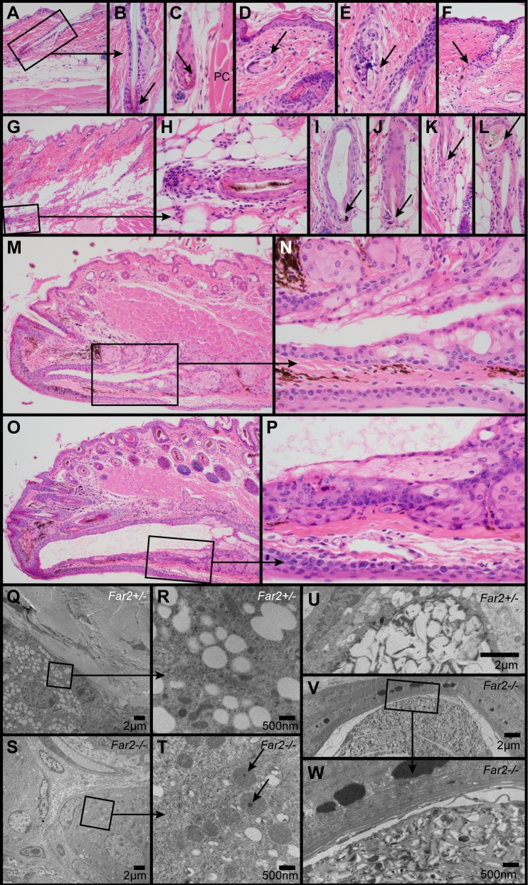
Fig 2. Histologic and ultrastructural changes in the skin.
Normal telogen follicle in a control mouse illustrates the normal sebaceous gland and club hair (arrow, A, B). In Far2-/- mice occasional telogen hair follicles (arrow) were present deep in the hypodermal fat layer adjacent to the panniculus carnosus (PC) muscle (C). Follicular dystrophy with or without rupture and associated inflammation was present in the dermis (arrow, D, E) and hypodermal fat layer (G-L). Occasional follicular scars (arrow, F) were present extending from below the sebaceous gland. Meibomian glands in the eyelids of wildtype mice (M, N) had a mildly dilated duct that was markedly dilated in Far2-/- mice (O, P). Ultrastructurally, a normal sebocyte in a female, 287 day old Far2+/- heterozygote mouse had numerous clear cytoplasmic vacuoles (Q). Higher magnification of boxed area (R) revealed the details of these vacuoles and surrounding organelles. By contrast, sebocytes in an age and sex matched Far2-/- homozygote mutant mouse had very few clear vacuoles and prominent oval mitochondria (S), some with electron dense material (arrow; T). A normal sebocyte in the sebaceous gland duct of a normal heterozygous female had numerous clear cytoplasmic vacuoles that were being compressed and ruptured (U). By contrast, sebocytes in an age and sex matched Far2-/- mouse had very few clear vacuoles and abundant cellular debris extending into the follicular infundibulum (V, W).

