Fig 1.
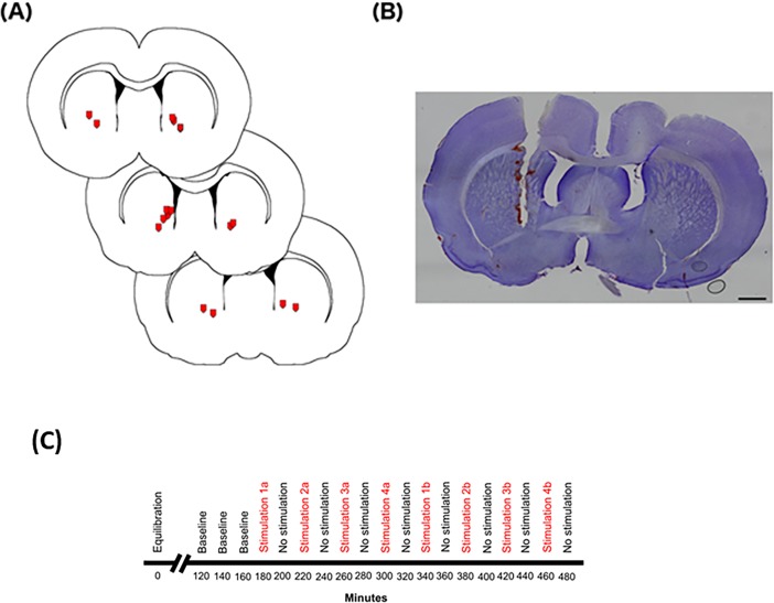
(A) Location of microdialysis probe tips the in striatum. Microdialysis probe locations (red symbols signify tip location) were seen between 0.50 mm and 1.20 mm from bregma based on Paxinos and Watson47. Probe membranes measure 2 mm upward from the tip of the probe. (B) Cresyl violet-stained tissue showing a microdialysis probe track through the cortex and into the striatum. Scale bar represents 1000 μm. (C) Timeline of microdialysis sampling. A 2 μm microdialysis probe was inserted into the striatum of urethane-anaesthetised rats. The timeline shows min following probe insertion. Artificial cerebrospinal fluid was perfused through the probe at a rate of 1.5 μl/min for the entirety of the sampling time. After 2 h of equilibration samples were collected every 20 min under different conditions.

