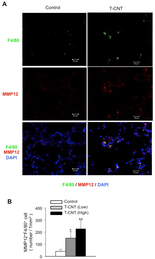
Fig 7. Detection of MMP-12-expressing alveolar macrophages in T-CNT-exposed mice.
(A) MMP-12 and F4/80 expression using frozen lung tissues of control and high-dose T-CNT-exposed mice were analyzed by confocal microscopy. Nuclei were stained with DAPI. Photos are representative of five mice of each group. (B) Number (mm2) of F4/80+MMP-12+ alveolar macrophages was counted. Data are presented as the average of relative expression to control ± SD of five mice of each group. *p < 0.05, **p < 0.005, vs controls.

