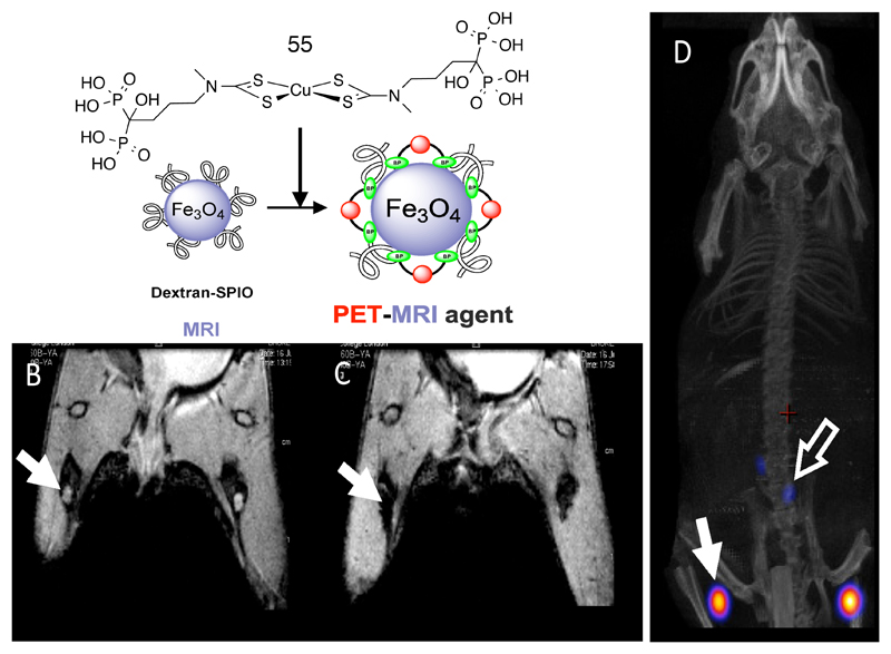
Figure 13.
Top: incorporation of 64Cu into iron oxide nanoparticles exploiting the affinity of copper complexes with pendant bisphosphonate groups (55) to the mineral surface. Bottom left: MRI images before (left) and after (right) injection of labeled particles into the footpads of a mouse, showing translocation of particles into lymph nodes indicated by arrows (as identified by loss of signal in the lymph nodes in the right image). On the right is a PET/CT image showing the location of 64Cu in the same lymph nodes as the MRI contrast.

