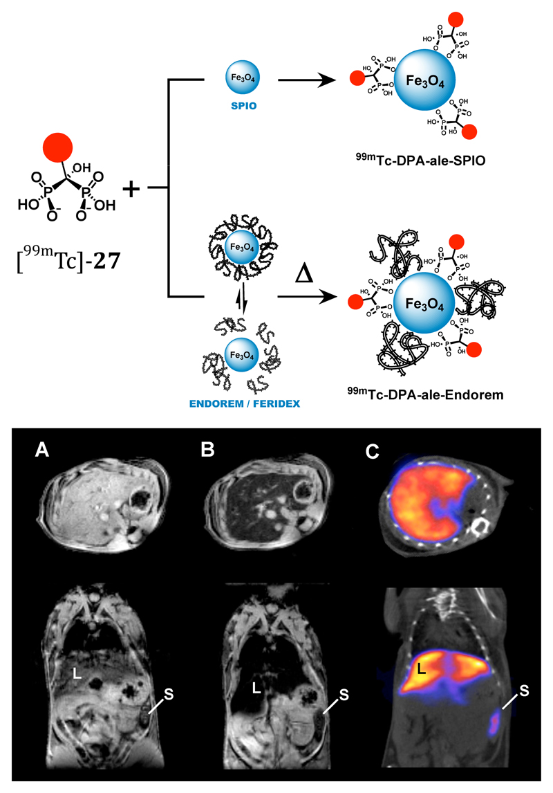
Figure 11.
Dual-modality (MRI/SPET) contrast agent combining iron oxide nanoparticles with 99mTc radiolabel linked to the inorganic iron oxide surface via bisphosphonate groups. Top: assembly of composite particle; bottom: Mouse images, transverse sections through liver. A: MR image pre-injection of contrast agent; B: MR image post-injection of contrast agent showing darkening of liver due to accumulation of iron oxide nanoparticles; C: SPET/CT image showing co-localisation of 99mTc with iron oxide contrast in liver (L) and spleen (S).

