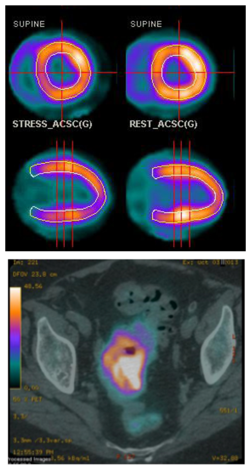
Figure 2.
Top: Example of myocardial perfusion PET imaging using 82Rb+ (transverse (top) and sagittal (bottom) sections); bottom: PET-CT scan (transverse section, pelvic region) of a patient with colorectal cancer after injection of 82Rb+. (Images courtesy of Prof A. M. Groves, University College London Hospitals)

