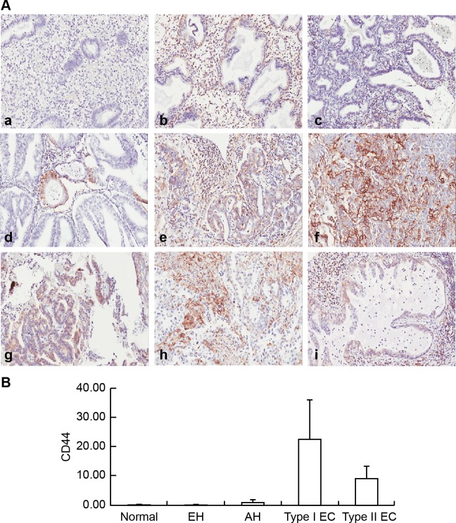
Fig 2. Examples of immunohistochemical staining for CD44 in endometrial lesions.
(A) Immunohistochemical examination of CD44 expression in normal endometrium (a), EH without atypia (b), AH(c), grade 1 EmAC (d), grade 2 EmAC (e), G3 EmAC (f), SC (g), CC (h), and MC (i). (B) Semiquantitative Comparison of CD44 Immunostaining Scores Between Normal Endometrium, EH without Atypia, AH, and Type I, and Type II EC. One-way ANOVA, p = 0.229.

