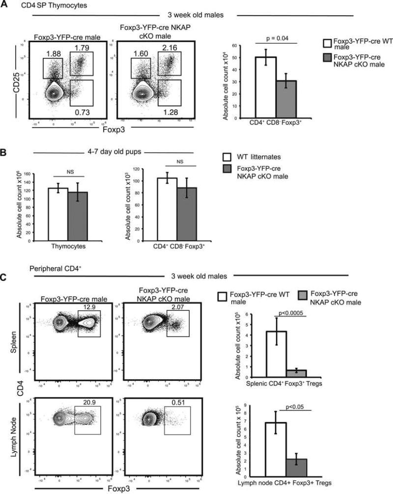
Figure 3. NKAP deficient Tregs develop in the thymus but fail to persist in the periphery.
(A) Examination of thymic development of Tregs at three weeks of age. Analysis of frequency of CD25+ Foxp3−, CD25− Foxp3+ and CD25+ Foxp3+ cells and absolute cell count of CD4+ CD8− Foxp3+ thymic Tregs. Data is from at least 6 mice per genotype from 3 independent experiments, and bar graphs show mean absolute cell count and SEM. (B) Examination of Treg development in pups aged 4–7 days. Data is from a total of 5 experiments with at least 5 mice in each genotype, and bar graphs show average absolute cell count and SEM. (C) Examination of Treg frequency and absolute cell counts in peripheral lymphoid organs (spleen, and pooled brachial, inguinal and cervical LN). Data is from at least 4 representative independent experiments with at least 4 mice per group, and bar graphs show mean absolute cell count and SEM.

