
Figure 3: Overexpression of miR-146a promotes cardiac dysfunction in vivo.
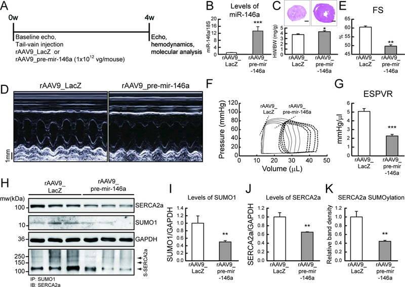
A, Protocol for AAV9-mediated pre-mir-146a gene transfer into normal mice. Six week-old male B6C3/F1 mice received AAV9 carrying pre-mir-146a (rAAV9_pre-mir-146a, 1×10¹² vg/mouse) or control virus (rAAV9_LacZ) via tail-vein injection. After 4 weeks of gene delivery, cardiac function was measured by echocardiography and then, further analyzed by hemodynamic measurements. B, levels of miR-146a. C, Cross-section images of a mouse heart (top) and quantification of heart weight/body weight ratio (bottom). Scale bars, 1 mm D and E, Representative LV M-mode images (D) and Factional shortening (FS) (E) (n=8). F and G, Representative pressure-volume loops (F) and ESPVR (G) (n=6). G, Representative blot showing SERCA2a, SUMO1 and total SUMOylation levels after infection with rAAV9_pre-mir-146a. I-K, The protein quantification of SUMO1 (I), SERCA2a (J) and SERCA2a SUMOylation (K) (n=6). *, p<0.05, **, p<0.01, ***, p<0.001 versus mouse infected with rAAV9_LacZ, as determined by Student’s t-test. Data are presented as mean ± s.e.m. in all panels.