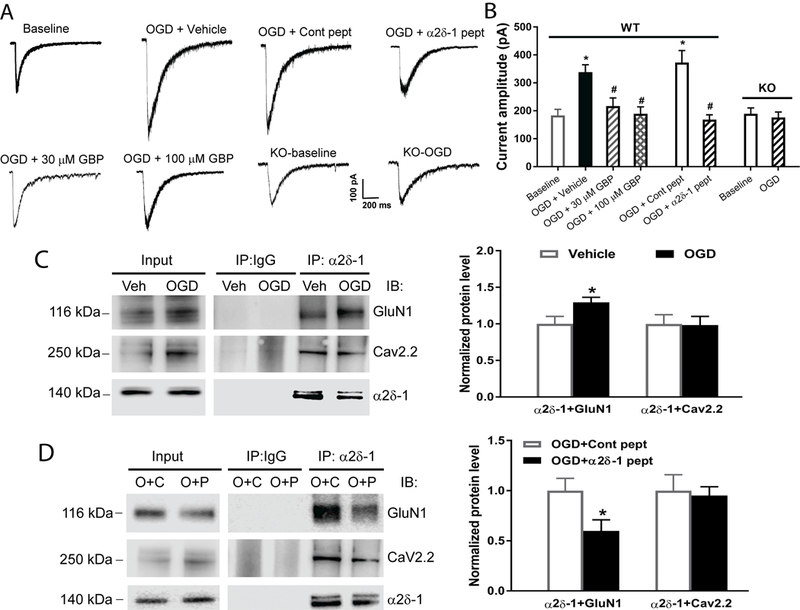
Figure 1. α2δ−1 and its interaction with NMDARs are essential for OGD-induced potentiation of NMDAR activity in hippocampal CA1 neurons.

(A and B) Representative recording traces (A) and quantification (B) of puff NMDA-elicited NMDAR currents show the effects of gabapentin (GBP, 30 µM for 60 min, n = 10 neurons; 100 µM for 60 min, n = 12 neurons), vehicle (n = 11 neurons), α2δ−1Tat peptide (1 µM for 60 min, n = 10 neurons), control peptide (cont peptide; 1 µM for 60 min, n = 10 neurons), and Cacna2d1 KO (n = 12 neurons for baseline and OGD groups) on NMDAR activity in hippocampal CA1 neurons subjected to 5 min of OGD. (C) Representative gel images and quantification of co-IP show the effect of OGD on the α2δ−1–GluN1 or α2δ−1–Cav2.2 association in mouse brain tissues (n = 6 mice per group). (D) Original gel images and quantification of co-IP show the effect of α2δ−1Tat peptide (P) and control peptide (C; both 1 µM for 30 min) on the α2δ−1–GluN1 or α2δ−1–Cav2.2 association in mouse brain slices subjected to OGD (O, n = 6 mice per group). Data are shown as means ± SEM. *p < 0.05 compared with the baseline current amplitude before OGD or with the vehicle or control peptide-treated group. #p < 0.05 compared with the OGD+vehicle or OGD+control peptide group.
