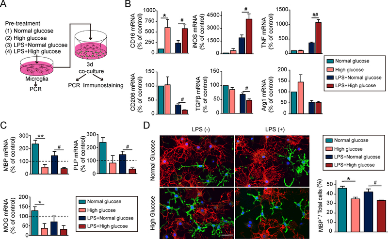
Figure 6. High glucose shifts microglial polarization toward M1, which impairs oligodendrocyte precursor cell differentiation under both physiological and inflammatory conditions.

A, Experimental design: Microglia in transwells were exposed to media with a normal concentration of glucose (5.5 mM) or high glucose (15 mM) for 48h in the presence or absence of a low concentration of LPS (2.5 ng/mL). Pretreated microglia were then co-cultured with oligodendrocyte precursor cells (OPCs) for 3d. B, The RNA expression of M1 and M2-like markers in microglia in transwells were measured by qRT-PCR. Expression of gapdh was used as an internal control. Data are fold-change relative to non-treated microglia. C, Expression of mbp, plp and mog in OPCs was assessed by qRT-PCR. Expression of gapdh was used as an internal control. Data are fold-change relative to OPCs without microglia co-cultures (dashed line). D, Double immunostaining for mature oligodendrocyte marker MBP (red) and OPC marker NG2 (green) in OPCs co-cultured for 3d with microglia-in-transwells. OPC differentiation was quantified by the percentage of MBP+ cells out of total cells (MBP++NG2+). Data were collected from 3 independent experiments. Scale bar: 50 μm. *p<0.05, **p<0.01, ***p<0.001; one way ANOVA and Bonferroni post hoc.
