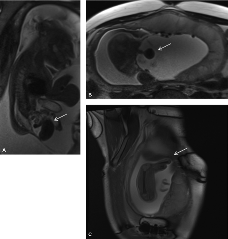
Fig. 2.

Case 2 sagittal ( A, C ) and axial ( B ) SSFSE MRIs. ( A, B ) No fluid-filled bladder, with “elephant trunk” midline loop of bowel and lateralized hemibladder masses. ( C ) Amputated lower limb in patient with myelomeningocele and nonvisualized bladder. MRI, magnetic resonance imaging; SSFSE, single-shot fast spin-echo.
