
Fig. 4.
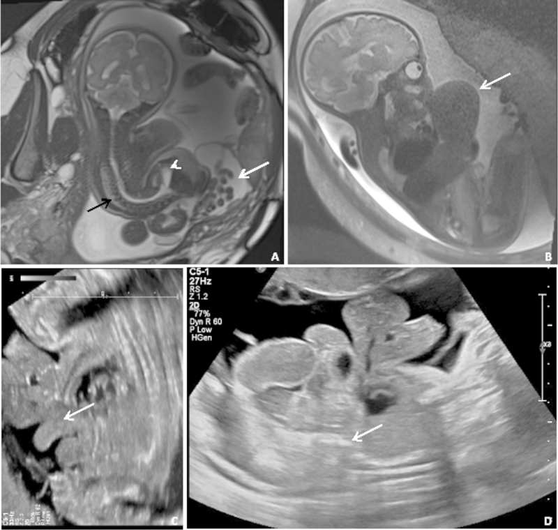
Coronal SSFP ( A ) and sagittal SSFSE ( B ) MRIs and two transabdominal ultrasound images of several older cases from our center. ( A ) Severe scoliosis (black arrow), abdominoschisis (white arrow), and meningocele (short arrow), but without body wall fusion. ( B ) Supraumbilical defect (white arrow) and absent bladder. ( C ) “Elephant trunk” sign representing prolapsed terminal ileum. ( D ) Fetal fusion to the placenta (white arrow) with large abdominoschisis. MRI, magnetic resonance imaging; SSFP, steady-state free precession; SSFSE, single-shot fast spin-echo.