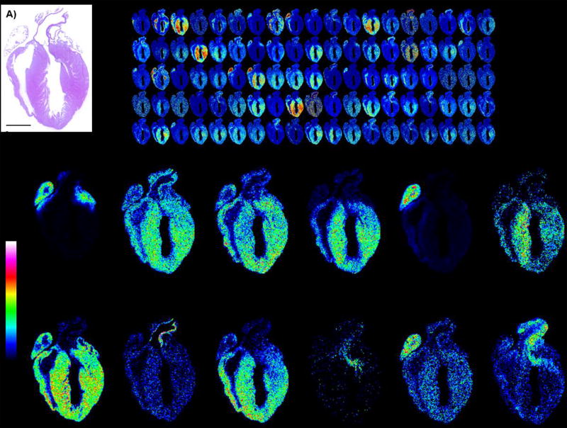
Figure 6.
Example images of expected output shown as MALDI IMS of tryptic peptides from mouse heart. A) H&E stain of heart. B) Example images of tryptic peptide expression showing 110 images out of 1,469 monoisotopic peaks (excluding isotopes 2 and 3 of isotopic envelopes). C) Example single images. Mouse heart tissue section donated by Dr. Christine Kern, Medical University of South Carolina. Mouse heart was fixed in 4% paraformaldehyde, paraffin embedded, and sectioned at 5 µm thickness.

