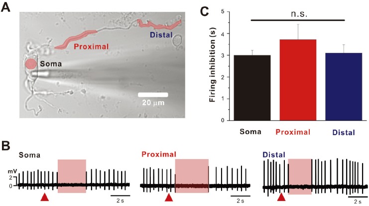
Fig. 4. Inhibition of spontaneous firing by caged-GABA uncaging on the soma and dendrites in DA neurons.
(A) Transmitted image of SNc DA neuron with uncaging sites of (O)-CNB caged-GABA (20 µM). (B) Inhibition of spontaneous firing by uncaging of caged-GABA on the soma, proximal dendrite, and distal dendrite, respectively. Red areas indicated the duration of firing inhibition by caged-GABA uncaging (red triangles) (C) Duration of spontaneous firing inhibition by local caged-GABA uncaging is summarized (n=5). n.s., nonsignificant.

