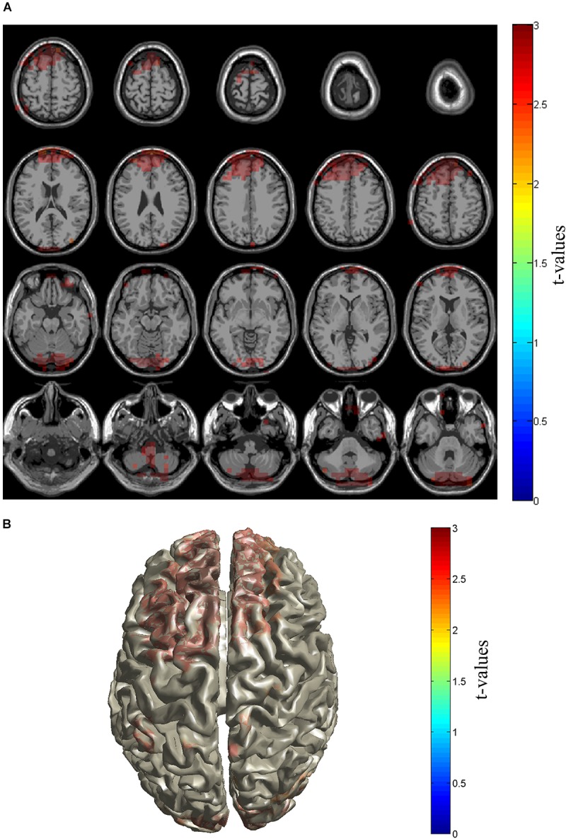
FIGURE 3.

Source distribution of gamma power. Sources with enhanced gamma power in PHN patients were shown in slice (A) and brain cortex (B). Sources locate in the prefrontal cortex including dorsolateral and medial prefrontal cortices as well as cerebellum area. The t-values are color-coded and only significant changes are displayed.
