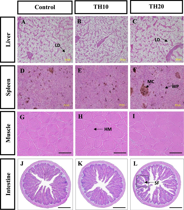
Figure 3.
Representative micrographs of liver, spleen, muscle and intestine of juvenile barramundi after 8 weeks of being fed with control, TH10 and TH20. (A–C) Liver histology from control (A) and TH20 (C) contain increased lipid deposition in hepatocytes while normal cells were observed in TH10 (B) fed fish. (D–F) Light micrographs of spleen showing marked melanomacrophage aggregates in TH20 (F) whereas such cases were not observed in control (D) and TH10 (E) diets. (G–I) Muscle tissues containing different diets showed healthy myotomes characterised by rounded, packed and uniformly identical muscle fibres. (J–L) The distal intestine of fish fed TH20 (L) showing reduced mucosal fold lengths and loss of epidermal integrity whereas control (J) and TH10 (K) fed fish intestinal fold were appear to be healthy with no obvious signs of intestinal inflammation. (LD = lipid droplet; MC = melanomacrophages complex; WP = white pulps; HM = healthy myotome; SF = short fold. All sections are stained with H&E. Scale bar, 50 μm.

