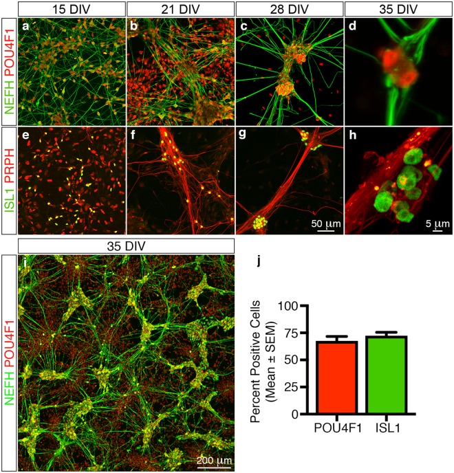
Figure 2.
Molecular characterisation of in vitro differentiated peripheral sensory neurons. Time-course analyses using established marker combinations demonstrate that the differentiated cells exhibit molecular signatures that are similar to in vivo peripheral sensory neurons. (a–d) Numerous POU4F1+/NEFH+ neurons are apparent in the differentiation cultures 1 day after replating (a, 15 DIV). The neurons then coalesce over time into POU4F1+ neuronal ganglia that radially project NEFH+ axonal tracts (b–d, 21 to 35 DIV). (e–h) Similarly, numerous ISL1+/PRPH+ neurons are also readily apparent in the differentiation cultures 1 day after replating (e, 15 DIV). These in turn also coalesce into ISL1+ neuronal ganglia that radially project PRPH+ axonal bundles (f–h, 21 to 35 DIV). (i) Overview image illustrating the elaborate in vitro neuronal networks that are composed of distinct POU4F1+ neuronal ganglia and radially projecting NEFH+ axonal tracts formed at 35 DIV. (j) Quantitative analysis of the differentiated neuronal cultures at 35 DIV demonstrates that 68 ± 4% and 72 ± 3% (mean ± SEM) of all neuronal ganglia contain POU4F1+ and ISL1+ cells, respectively. All images and quantification analyses were performed on neurons derived from at least three independent differentiation experiments. Abbreviations: DIV, days in vitro. Scale bars: (a–c, e–g) 50 μm, (d, h) 5 μm; (i) 200 μm.

