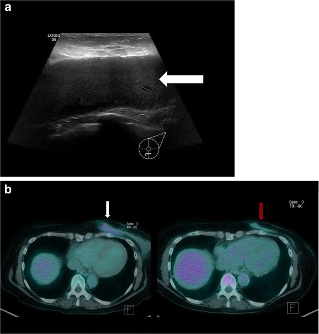
Fig. 2.
a Ultrasound revealed a large effusion with no signs of infection (white arrow). Fortunately, the aspirated fluid was sent for cytology, which confirmed BIA-ALCL. b PET/CT revealed a flattened rim of soft tissue, located inferomedially in the left breast, with ill-defined margins and moderate FDG uptake (white arrow). The patient subsequently received six cycles of chemotherapy and targeted radiotherapy. Restaging PET/CT revealed complete metabolic response (red arrow)

