
Fig. 13.
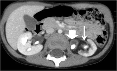
Diffuse XPN. Contrast-enhanced nephrogenic phase transverse CT shows bilateral kidney lithiasis. Right kidney presents a dilated pelvis with a stone inside, while left kidney shows a stone in the pelvis with fatty proliferation (thin white arrow) and an obstructive stone inside a dilated ureter (thick white arrow)