
Fig. 2.
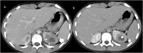
Focal XPN. Same patient as in Fig. 1. a Contrast-enhanced nephrogenic phase transverse CT image shows focal hypodense mass with rim enhancement (white arrows) and adjacent parenchymal destruction (discontinuous white arrow) in the cortex of the left kidney. Perirenal infiltration (thick white arrow) is present. b Contrast-enhanced excretory phase transverse CT image: the lesion is better delimited with thick rim enhancement and hypodense centre (asterisk). Working kidneys with contrast material excretion in the collecting system (white arrows) are shown. Perirenal infiltration (thick white arrow) is present