
Fig. 3.
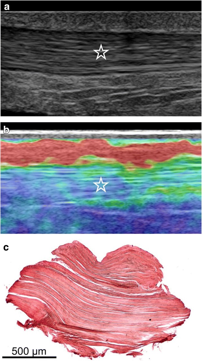
Images of a normal Achilles tendon using CE and histological correlation. a Conventional ultrasound (US) image of the middle portion of the Achilles tendon in the longitudinal plane. The star indicates the homogenous fibrillar pattern defining normal tendon appearance. b Image of ultrasound elastography (USE) at the same level as in a. The blue-green area of the elastogram represented by the star indicates tissue stiffness where biopsy was subsequently performed. c Histological image obtained with orcein staining showing parallel collagen fibrils, without adipose infiltration and capillary proliferation. Reproduced, with permission, from Klauser et al. [31], copyright (2013) by the Radiological Society of North America, Inc. (RSNA)