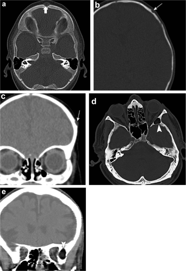
Fig. 14.
Dermoid cysts. Patient 1: Axial head CT (a) depicts a midline fat-containing lesion in the frontal region (thick arrow). Patient 2: Axial bone window (b) and coronal soft tissue window head CT (c) show a dermoid cyst in the left frontal bone involving the outer table (dashed arrows). Patient 3: Axial CT (d) and coronal head CT portray a fat-containing lesion in the left grater wing of the sphenoid (arrowheads)

