Fig. 23.
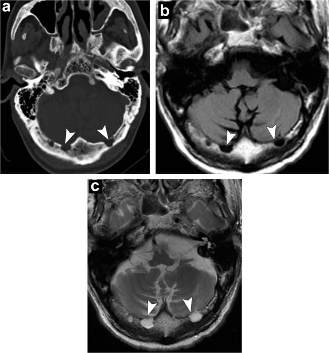
Arachnoid granulations. Axial head CT (a) shows well-defined structures along the inner table in the region of the transverse sinus (arrowheads). Axial T1-weighted (b) and axial T2-weighted (c) images demonstrate fluid signal of these structures following CSF along the transverse sinus extending into the occipital bone (arrowheads)

