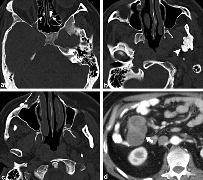
Fig. 4.
Gardner Syndrome (multiple osteomas). Axial head CT images (a–c) show multiple osteomas in the ethmoid air cells (dashed arrows), left ramus of the mandible (arrowhead) and right anterior wall of the maxillary sinus (thin arrow). This patient was also found to have a mesenteric desmoid tumour (thick arrow, d)

