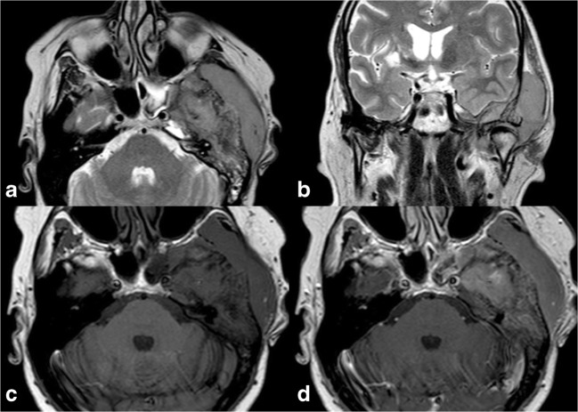
Fig. 12.
Primary lymphoma of the bone, same patient shown in Fig. 11. On MRI, the lesion demonstrates low T2 signal on axial (a) and coronal (b) T2-weighted sequences and extension to the temporal fossa and to the epidural space of the middle cranial fossa. The tumour exhibits homogeneous contrast enhancement; axial T1-weighted before (c) and after (d) contrast administration

