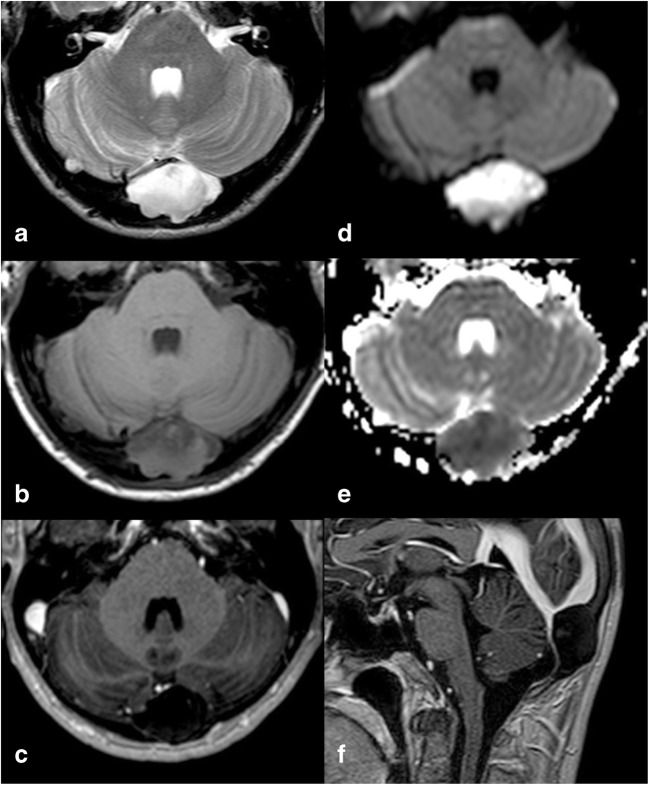
Fig. 4.
IEC. On MR images of the same patient shown in Fig. 3, the lesion appears slightly inhomogeneous, mostly hyperintense on T2w (a) and hypointense on T1w (b) images, without contrast enhancement (c), showing diffusion restriction on b 1000 diffusion-weighted imaging (DWI) and the apparent diffusion coefficient (ADC) map (d, e). It causes compression on the confluence of sinuses (sagittal contrast-enhanced T1w image, f). As a consequence, the patient presented with intracranial hypertension symptoms

