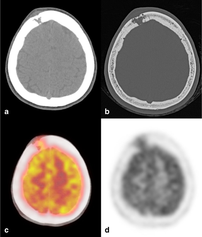
Fig. 9.
Eosinophilic granuloma (EG). Axial CT images with soft tissues (a) and bone (b) windows showing a right frontal lytic lesion with ill-defined margins extending in the contiguous extracranial soft tissues. Erosion of the inner cranial table is more pronounced than of the outer (“hole within a hole” sign). 18(F) FDG-PET CT examination demonstrates a moderate tracer uptake (c, d)

