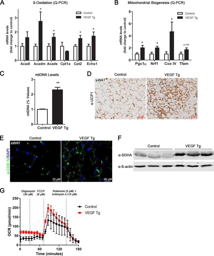
FIG 2.
Local overexpression of VEGF-A stimulates mitochondrial biogenesis and functions in adipose tissues. (A) qPCR analysis of β-oxidation-related genes, namely, Acadl, Acadm, Acads, Cpt1a, Cpt2, and Echs1, in sWAT of VEGF Tg mice and their littermate controls after HFD-Dox feeding for 7 days (n = 5 per group; Student's t test, *, P < 0.05). (B) qPCR analysis of mitochondrial biogenetic genes including the Pgc1α, Nrf1, CoxIV, and Tfam genes in sWAT of VEGF Tg mice and their littermate controls after HFD-Dox feeding for 7 days (n = 5 per group; Student's t test, *, P < 0.05). (C) The mitochondrial DNA (mtDNA) content in sWAT of VEGF Tg mice and their littermate controls after HFD-Dox feeding for 7 days. The copy number of mtDNA was calculated by the ratio of the mtDNA gene for NADH dehydrogenase alpha 1 (Nadha1) to the nuclear gene for lipoprotein lipase (Lpl) (n = 5 per group; Student's t test, **, P < 0.01). (D) IHC staining with anti-UCP1 antibody in sWAT of VEGF Tg mice and their littermate controls after HFD-Dox feeding for 7 days (scale bar, 50 μm). (E) Immunofluorescence (IF) staining with anti-Cox IV antibody (green) in sWAT of VEGF Tg mice and their littermate controls after HFD-Dox feeding for 7 days. The nuclei were stained with DAPI (blue). (F) Western blotting of protein levels of SDHA with anti-SDHA antibody in sWAT of VEGF Tg mice and their littermate controls after HFD-Dox feeding for 7 days. Equal loading control was demonstrated by anti-β-actin antibody (n = 3 per group). (G) Oxygen consumption rate (OCR) in sWAT measured by a Seahorse XFe24 instrument. The sWAT was collected from VEGF Tg mice and their littermate controls after HFD-Dox feeding for 7 days. The injection time point and final concentration of different compounds used for mitochondrial stress assay are indicated in the panel (n = 5 per group).

