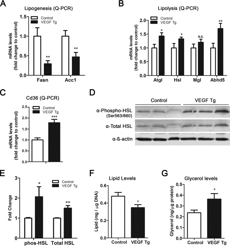
FIG 3.
Local overexpression of VEGF-A stimulates lipolysis in sWAT. (A) qPCR analysis of lipogenic genes, namely, Fasn and AccI, in sWAT of VEGF Tg mice and their littermate controls after HFD-Dox feeding for 7 days (n = 6 per group; Student's t test, **, P < 0.01). (B) qPCR analysis of lipolytic genes, namely, Atgl, Hsl, Mgl, and Abhd5, in sWAT of VEGF Tg mice and their littermate controls after HFD-Dox feeding for 7 days (n = 6 per group; Student's t test, *, P < 0.05; **, P < 0.01, N.S., not significant). (C) qPCR analysis of the Cd36 gene in sWAT of VEGF Tg mice and their littermate controls after HFD-Dox feeding for 7 days (n = 6 per group; Student's t test, ***, P < 0.001). (D and E) Western blotting and quantitative measurement of band density on the Western blots by ImageJ software for phosphorylated HSL (phos-HSL) at serine 563/660 as well as total HSL levels in sWAT of VEGF Tg mice and their littermate controls after HFD-Dox feeding for 7 days. Results were normalized by β-actin (n = 3 per group; Student's t test, *, P < 0.05; **, P < 0.01). (F) Total triglyceride content in sWAT of VEGF Tg mice and their littermate controls after HFD-Dox feeding for 7 days. (n = 4 per group; Student's t test, *, P < 0.05). (G) Glycerol levels (as the product of lipolysis) in sWAT of VEGF Tg mice and their littermate controls after HFD-Dox feeding for 7 days (n = 4 per group; Student's t test, *, P < 0.05).

