FIG 7.
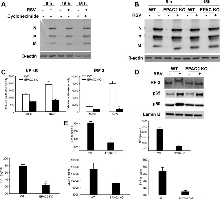
EPAC2 directly regulates RSV-induced host immunity. (A) Limited RSV protein accumulation along the course of infection. WT MEF cells were mock infected or infected with RSV at MOI of 1. At 2 h p.i., 20 μM cycloheximide was added as indicated. Untreated cells were used as controls. At 6 or 15 h p.i., total cell lysates were prepared and subjected to Western blotting using an anti-RSV antibody. β-Actin was used as a control for the efficacy of cycloheximide treatment. (B) RSV replication in wild-type (WT) or EPAC2 knockout (KO) MEFs. MEFs, with or without EPAC2, were mock infected or infected with RSV at an MOI of 1. At 6 or 15 h p.i., total cell lysates were harvested and subjected to Western blotting using an anti-RSV antibody. β-Actin was used as a control for equal loading of the samples. Data shown are representative of three independent experiments. (C) The impact of EPAC2 on the activation of NF-κB and IRF-3. WT or EPAC2 KO cells in triplicate were transfected with a luciferase reporter plasmid containing the human NF-κB (left) or IRF-3 (right) promoter. At 30 h posttransfection, cells were mock infected or infected with RSV for 15 h and harvested to measure luciferase activities. **, P < 0.01 relative to the RSV-infected WT MEFs. (D) EPAC2-controlled nuclear translation of NF-κB and IRF-3. WT or EPAC2 KO cells were mock infected or infected with RSV. At 15 h p.i., nuclear extracts of cells were prepared and subjected to Western blotting using an anti-IRF-3, anti-p65, or anti-p50 antibody. Lamin B was used as a control for equal loading of the samples. Data shown are representative of two independent experiments. (E) EPAC2-regulated cytokine/chemokine induction. WT and EPAC2 KO cells were mock infected or infected with RSV for 15 h, and supernatant was collected to measure cytokines/chemokines. Net induction data shown are representative of three independent experiments and means ± SE. **, P < 0.01 relative to the WT.

