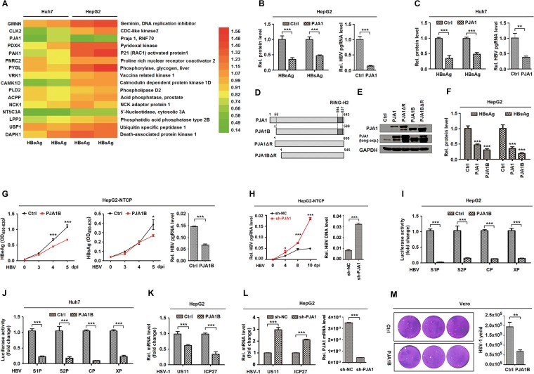
FIG 1.
PJA1 represses the transcription and replication of HBV and HSV-1. (A) Huh7 and HepG2 cells were plated in 24-well plates and transfected with 0.2 μg pHBV1.3 and 0.3 μg plasmids expressing 16 candidate proteins in triplicates for 48 h. (B and C) HepG2 cells (B) and Huh7 cells (C) were plated in 24-well plates and transfected with 0.2 μg pHBV1.3 and 0.3 μg pCAGGS-HA-PJA1 in triplicates for 48 h. HBeAg and HBsAg in the supernatants were assayed by an ELISA, and HBV pgRNA was measured by qPCR. (D) Diagrams of PJA1, PJA1B, PJA1ΔR, and PJA1BΔR proteins. (E) 293T cells were plated in 12-well plates and transfected with 1 μg pCAGGS-HA, pCAGGS-HA-PJA1, pCAGGS-HA-PJA1ΔR, pCAGGS-HA-PJA1B, and pCAGGS-HA-PJA1BΔR for 48 h. The expressed proteins were detected by Western blotting using anti-PJA1 antibody. (F) HepG2 cells were plated in 24-well plates overnight and transfected with 0.2 μg pHBV1.3 and 0.3 μg pCAGGS-HA, pCAGGS-HA-PJA1, and pCAGGS-HA-PJA1B for 48 h. HBeAg and HBsAg in the supernatants were assayed by an ELISA. (G) HepG2-NTCP cells were plated in 6-well plates, transfected with 2 μg pCAGGS-HA or pCAGGS-HA-PJA1B, and infected with HBV at 1,000 GE by inoculation with concentrated supernatants of HepaAD38 cells. HBeAg and HBsAg in the supernatants were assayed by an ELISA, and HBV pgRNA was measured by qPCR. OD450-630, optical density at 450 to 630 nm. (H) HepG2-NTCP cells were plated in 6-well plates, transfected with 2 μg pLKO.1-sh-NC or -sh-PJA1 for 24 h, and infected with HBV at 1,000 GE by inoculation with concentrated supernatants of HepaAD38 cells. sh-NC, nonspecific control shRNA. HBV pgRNA was measured by qPCR. Total DNA was extracted at 12 days postinfection (dpi), and DNA levels of HBV replication intermediates were measured by qPCR. (I and J) HepG2 cells (I) and Huh7 cells (J) were plated in 24-well plates and transfected with 0.3 μg pCAGGS-HA-PJA1B and 0.2 μg luciferase reporters containing the pre-S1, pre-S2, core, and X promoters of HBV for 48 h. Luciferase activities were determined, and results are expressed as fold induction relative to the control. (K) HepG2 cell lines stably expressing PJA1B were generated and infected with HSV-1 at an MOI of 0.1 for 8 h. HSV-1 US11 and ICP27 mRNA levels were determined by RT-qPCR. (L) HepG2-sh-NC and HepG2-sh-PJA1 cells were infected with HSV-1 at an MOI of 0.1 for 8 h. (Left) HSV-1 US11 and ICP27 mRNA levels were determined by RT-qPCR. (Right) HepG2 cell lines stably expressing pLKO.1-sh-NC or -sh-PJA1 were generated, and PJA1 mRNA levels in HepG2-sh-NC and HepG2-sh-PJA1 cells were detected. (M) Vero cells were plated in 6-well plates, transfected with 2 μg pCAGGS-HA or pCAGGS-HA-PJA1B for 24 h, and infected with HSV-1 at an MOI of 0.1. At 48 h postinfection, cell culture supernatants were collected, and the viral yields were determined by a plaque assay. Data are shown as means ± SD and correspond to results from a representative experiment out of three performed. **, P < 0.01; ***, P < 0.001.

