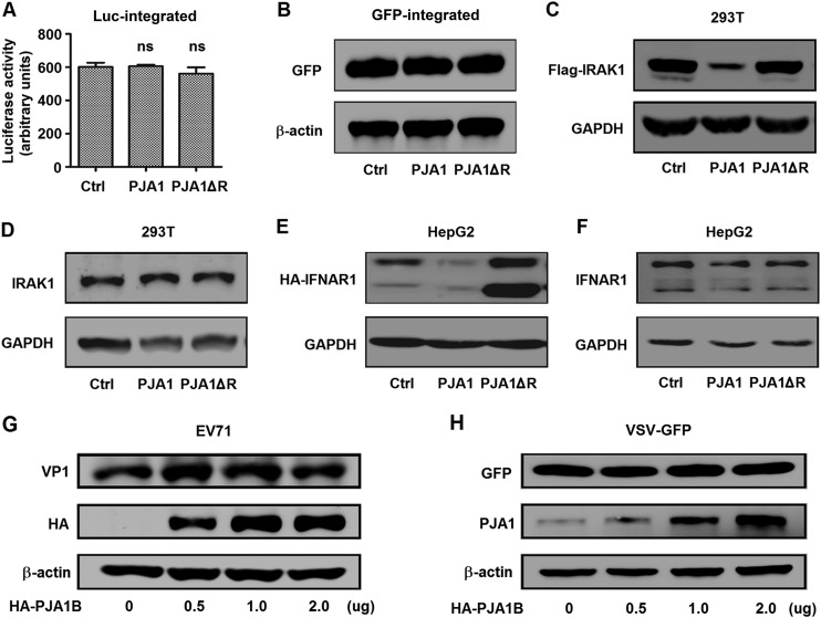
FIG 3.
PJA1 has no effect on chromosome-integrated genes and RNA viruses. (A) 293T-Luc cells were generated, in which the CMV promoter driving Luc was randomly integrated into cell chromosomes by a lentiviral system. The cells were plated in 24-well plates and transfected with 0.5 μg pCAGGS, pCAGGS-HA-PJA1, or pCAGGS-HA-PJA1ΔR for 48 h. Luciferase activities were measured. Data are shown as means ± SD and correspond to results of a representative experiment out of three performed. ns, nonsignificant. (B) 293T-EGFP cells were generated, in which the CMV promoter driving EGFP was randomly integrated into cellular chromosomes by a lentiviral system. The cells were plated in 12-well plates and transfected with 1 μg pCAGGS, pCAGGS-HA-PJA1, or pCAGGS-HA-PJA1ΔR. GFP and β-actin protein were detected by Western blot analyses. (C) 293T cells were plated in 12-well plates and cotransfected with 0.5 μg pFlag-IRAK1 and 0.5 μg pCAGGS, pCAGGS-HA-PJA1, or pCAGGS-HA-PJA1ΔR for 48 h. Flag-IRAK1 and GAPDH protein levels were determined by Western blot analyses. (D) 293T cells were plated in 12-well plates and transfected with 1 μg pCAGGS, pCAGGS-HA-PJA1, or pCAGGS-HA-PJA1ΔR for 48 h. Endogenous IRAK1 and GAPDH were detected by Western blot analyses. (E) HepG2 cells were plated in 12-well plates and cotransfected with 0.5 μg pCAGGS-HA-IFNAR1 and 0.5 μg pcDNA3.1, pcDNA3.1-PJA1, or pcDNA3.1-PJA1ΔR for 48 h. HA-IFNAR1 and GAPDH were detected by Western blot analyses. (F) HepG2 cells were plated in 12-well plates and transfected with 1 μg pcDNA3.1, pcDNA3.1-PJA1, or pcDNA3.1-PJA1ΔR for 48 h. Endogenous IFNAR1 and GAPDH were detected by Western blot analyses. (G) RD cells were plated in 6-well plates, transfected with pCAGGS-HA-PJA1B at different concentrations (0, 0.5, 1, and 2 μg) for 48 h, and infected with EV71 at an MOI of 5 for 6 h. EV71 VP1, HA-PJA1B, and GAPDH were detected by Western blot analyses. (H) 293T cells were plated in 6-well plates, transfected with pCAGGS-HA-PJA1B at different concentrations (0, 0.5, 1, and 2 μg) for 24 h, and infected with VSV-GFP at an MOI of 1 for 12 h. GFP, PJA1B, and GAPDH were detected by Western blot analyses.

