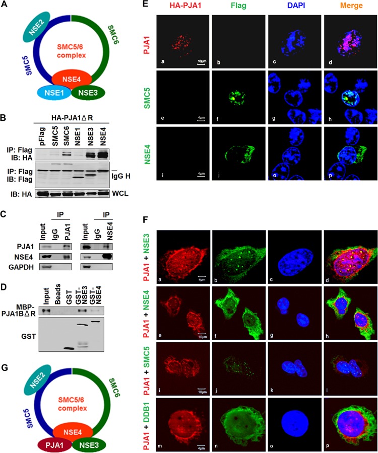
FIG 5.
PJA1 interacts with the SMC5/6 complex in the nucleus. (A) Schematic of the SMC5/6 complex. (B) 293T cells were plated in 6-well plates and cotransfected with 1 μg pCAGGS-HA-PJA1ΔR and 1 μg pFlag, pFlag-SMC5, pFlag-SMC6, pFlag-NSE1, pFlag-NSE3, or pFlag-NSE4. Cells were lysed in RIPA lysis buffer. The immunoprecipitates and whole-cell lysates were analyzed by Western blotting with anti-HA or anti-Flag antibody. IB, immunoblot. (C) 293T cells were lysed in RIPA lysis buffer, and cell lysates were immunoprecipitated with anti-PJA1 antibody, anti-NSE4 antibody, or rabbit IgG. The immunoprecipitates and whole-cell lysates were analyzed by Western blotting with antibody to PJA1, NSE4, or GAPDH. (D) GST pulldown with cell lysates containing MBP-PJA1BΔR and GST, GST-NSE3, or GST-NSE4 expressed and purified from E. coli. After pulldown, precipitates were analyzed by Western blotting with anti-PJA1 or anti-GST antibody. (E and F) HepG2 cells were plated in confocal dishes and transfected with 1 μg pCAGGS-HA-PJA1, pFlag-SMC5, or pFlag-NSE4 (E) or cotransfected with 1 μg pCAGGS-HA-PJA1 and 1 μg pFlag-SMC5, pFlag-NSE3, pFlag-NSE4, or pFlag-DDB1 (F). Cells were immunostained with anti-HA and anti-Flag antibodies. Immunofluorescence analysis shows PJA1 (red), NSE3/4 (green), SMC5 (green), and DDB1 (green). The nucleus was stained by DAPI. (G) Schematic of the SMC5/6 complex in which NSE1 was replaced by PJA1.

