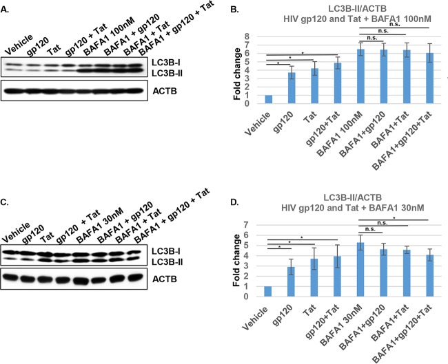
FIG 10.
Treatment with gp120 and Tat induces blockage of mitophagic flux in neurons. (A to D) Protein expression levels of LC3B-II were analyzed by immunoblotting with LC3B antibody in neuronal cell lysates treated with HIV gp120, Tat, or both for 24 h and with 100 nM (A) or 30 nM bafilomycin A1 (BAFA1) (C) for 8 h prior to collection. Beta-actin (ACTB) was used as an internal loading control. (B, D) The relative expression of LC3B-II was normalized to the expression of ACTB. Each data point was normalized to the results for treatment with vehicle and analyzed by Image J software. Student's t test was performed to test for statistical significance. Data are presented as mean values ± SD (n = 3 independent donors). *, P < 0.05; n.s., not significant.

