FIG 2.
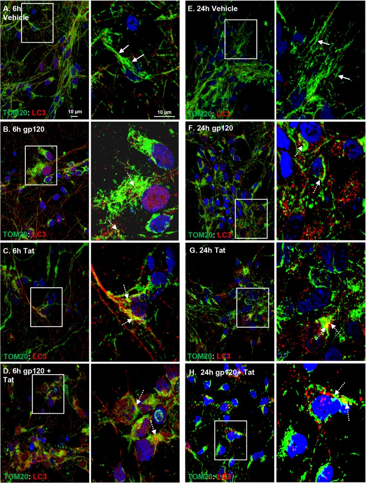
HIV gp120 and Tat induce mitochondrial fission and mitophagosome formation. Confocal laser microscopy analysis showing a healthy tubular network of mitochondria in vehicle-treated cells (A, E) and mitochondrial fragmentation in HIV gp120- and Tat-treated neurons (B to D, F to H) 6 h posttreatment with 100 ng/ml HIV gp120 (B), Tat (C), or both (D) and 24 h posttreatment with 100 ng/ml HIV gp120 (F), Tat (G), or both (H). Neurons were immunostained with TOM20 mitochondrial marker (green) and LC3B-II autophagosome marker (red). In the enlarged images of boxed areas, solid white arrows indicate typical tubular healthy mitochondria and dashed white arrows indicate fragmented mitochondria. Colocalization of mitochondria with LC3 autophagosomes (mitophagosomes) appears yellow. Scale bars, 10 μm.

