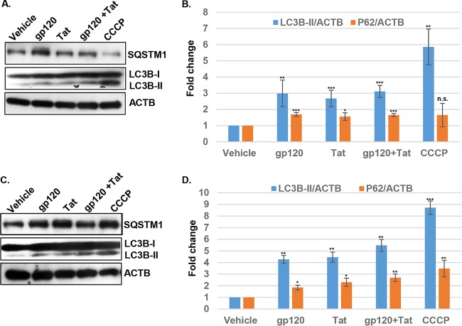
FIG 3.
HIV gp120 and Tat increase LC3II lipidation and P62 expression 6 h posttreatment with 100 ng/ml HIV gp120, Tat, or both (A) and 24 h posttreatment with 100 ng/ml HIV gp120, Tat or both (C). CCCP was used as the positive control. Neuronal cell lysates were extracted with mitochondrial lysis buffer, clarified by centrifugation, and analyzed by Western blotting using antibodies against LC3B and SQSTM1. Beta-actin (ACTB) was used as an internal loading control. (B and D) The relative expression of LC3B-II and SQSTM1 (P62) was normalized to that of beta-actin. Each data point was normalized to the corresponding result for vehicle-treated cells and analyzed by Image J software. Student's t test was performed to test for statistical significance. Data are presented as mean values ± standard deviations (SD) (n = 3 independent donors). *, P < 0.05; **, P < 0.01; ***, P < 0.001; n.s., not significant.

